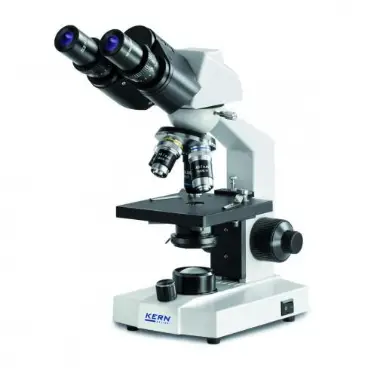
What Is A Light Microscope?
Microscopes are very important tools for scientists; they are used in microbiology, material science, mineralogy and medicine. A light microscope can simply be defined as, an instrument that uses visible light and magnifying lenses to examine small objects not visible to the naked eye or in finer detail than the naked eye allows. Light microscopes send light through a path that first focuses the light into a tight beam and then passes that light through a sample, which creates an image. That image then passes one or more lenses to magnify it until it reaches the user’s eye or camera.
What You Need To Know About Light Microscope
- Light microscope uses visible light to illuminate the specimen.
- Preparation of specimen to be viewed under a light microscope takes a few minutes to hours.
- The size of a light microscope is relatively smaller and can be operated as a desktop instrument.
- Light microscope condenser, objective lens and eye piece lenses are made of glass.
- In light microscope, the image formation depends upon light absorption in different zones of the object.
- In light microscope, image is seen by eyes through ocular lenses (eye piece). No screen is needed.
- Light microscope has a magnification of 500X to 1500X.
- Fixed or unfixed, stained or unstained living or non-living specimen can be observed under a light microscope.
- While using a light microscope, specimen is usually mounted on glass slide
- When using a light microscope, magnification is changed by changing the objective or eye piece lenses.
- When using a Light microscope, color imparting dyes are used for staining to provide contrast and differentiation.
- In light microscope, live cell imaging is possible and hence the living cellular processes can be visualized.
- It is possible to visualize the natural color of the specimen under a light microscope.
- Vacuum condition is not required for the working of a light microscope.
- Light microscope has a relatively low resolving power of about 200 nm.
- A light microscope does not use filaments anywhere in its operation.
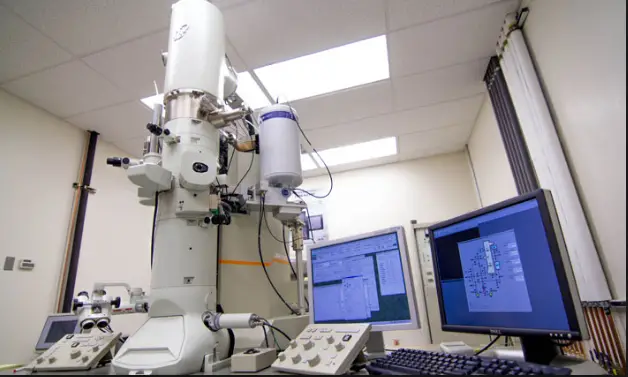
What Is An Electron Microscope?
An electron microscope can simply be defined as a microscope that focuses beams of energetic electrons to examine objects up to nano-scales. It works by using an electron beam instead of visible light and an electron detector instead of our eyes. Electron microscopes have much greater resolving than light microscopes and can obtain much higher magnification. Both electron and light microscopes have resolution limitations imposed by their wavelength.
What You Need To Know About Electron Microscope
- Electron microscope uses a beam of electrons (radiations) to illuminate the specimen.
- Preparation of specimen to be viewed under electron microscope often takes several days.
- The size of an electron microscope is relatively larger due to separate systems such as cooling system, image processing system, vacuum system etc.
- In electron microscope, all lenses are electromagnetic.
- In electron microscope, image formation depends upon the scattering of electron beams by different regions of the object due to heavy metal staining.
- In electron microscope the image is received on zinc sulphate fluorescent screen or photographic plate.
- Electron microscope has a magnification of 100000X to 500000X.
- Only fixed and stained non-living specimen can be observed under an electron microscope.
- When using an electron microscope, specimen is mounted on metallic grid (usually copper).
- When using an electron microscope, magnification is changed by adjusting the power of electric current to the electromagnetic lenses.
- In electron microscope, heavy metals are used as stains, which deflect the electron rays to produce the image.
- While using an electron microscope, live cell imaging is not possible and hence the living cellular processes cannot be visualized.
- It is not possible to visualize the natural color of specimen under electron microscope.
- Vacuum condition is essential for its working because the electron beams have a shorter wave length and can easily be destroyed or deflected by molecules in the air.
- An electron microscope has a resolving power of 0.1 nm.
- An electron microscope uses tungsten filament to produce electrons.
The Difference Between Light Microscope And Electron Microscope In Tabular Form
| Basis of Comparison | Light Microscope | Electron Microscope |
| Illuminating Source | Uses visible light to illuminate the specimen. | Uses a beam of electrons (radiations) to illuminate the specimen. |
| Specimen Preparation | Preparation of specimen to be viewed under a light microscope takes a few minutes to hours. | Preparation of specimen to be viewed under electron microscope often takes several days. |
| Size of the Instrument | The size of a light microscope is relatively smaller and can be operated as a desktop instrument. | The size of an electron microscope is relatively larger due to separate systems such as cooling system, image processing system, vacuum system etc. |
| Lens | Condenser, objective lens and eye piece lenses are made of glass. | All lenses are electromagnetic. |
| Image Formation | The image formation depends upon light absorption in different zones of the object. | Image formation depends upon the scattering of electron beams by different regions of the object due to heavy metal staining. |
| Image View | Image is seen by eyes through ocular lenses (eye piece). No screen is needed. | The image is received on zinc sulphate fluorescent screen or photographic plate. |
| Magnification | Has a magnification of 500X to 1500X. | Has a magnification of 100000X to 500000X. |
| Nature of Specimen | Fixed, unfixed, stained or unstained living or non-living specimen can be observed under a light microscope. | Only fixed and stained non-living specimen can be observed under an electron microscope. |
| Specimen Mounting | Specimen is usually mounted on glass slide. | Specimen is mounted on metallic grid (usually copper). |
| How Magnification is changed | Magnification is changed by changing the objective or eye piece lenses. | Magnification is changed by adjusting the power of electric current to the electromagnetic lenses. |
| Stains | Color imparting dyes are used for staining to provide contrast and differentiation. | Heavy metals are used as stains, which deflect the electron rays to produce the image. |
| Limitation | Live cell imaging is possible and hence the living cellular processes can be visualized | Live cell imaging is not possible and hence the living cellular processes cannot be visualized. |
| Color Visualization | It is possible to visualize the natural color of the specimen under a light microscope. | It is not possible to visualize the natural color of specimen under electron microscope. |
| Vacuum Condition | Vacuum condition is not required for the working of a light microscope. | Vacuum condition is essential for its working because the electron beams have a shorter wave length and can easily be destroyed or deflected by molecules in the air. |
| Resolving Power | Has a relatively low resolving power of about 200 nm. | Has a resolving power of 0.1 nm. |
| Filament | Does not use filaments anywhere in its operation. | Uses tungsten filament to produce electrons. |
Advantages Of Electron Microscope Over Light Microscope
- Has ability to produce powerful magnification.
- It offers a higher resolution than what is possible with light microscope.
- Has diverse range of applications in technology, for example it is heavily used in manufacturing of computer chips and quality control assurance testing.
- Electron microscopes such as scanning electron microscope have ability to view hydrated specimens in low-pressure or wet environments.
Summary
What Is The Main Difference Between Light Microscope And An Electron Microscope?
Light microscope uses visible light to illuminate the specimen while the electron microscope uses a beam of electrons (radiations) to illuminate the specimen.